Pelvic ultrasound (transvaginal): Difference between revisions
No edit summary |
|||
| (14 intermediate revisions by 2 users not shown) | |||
| Line 1: | Line 1: | ||
==Background== | ==Background== | ||
[[File:Blausen 0732 PID-Sites.png|thumb|Pelvic anatomy.]] | |||
*Also known as endovaginal ultrasound | *Also known as endovaginal ultrasound | ||
*Used to evaluate female reproductive organs including uterus, ovaries, fallopian tubes, cervix, and vagina | *Used to evaluate female reproductive organs including uterus, ovaries, fallopian tubes, cervix, and vagina | ||
==Indications== | ==Indications== | ||
===General=== | ===General=== | ||
*Pelvic pain | *[[Pelvic pain]] | ||
*Abnormal pelvic or abdominal exam | *Abnormal pelvic or abdominal exam | ||
| Line 20: | Line 21: | ||
*Hydrosalpinx | *Hydrosalpinx | ||
===Pregnancy=== | ===[[Pregnancy]]=== | ||
*Confirm intrauterine pregnancy | *Confirm intrauterine pregnancy | ||
*Gestational dating | *Gestational dating | ||
| Line 82: | Line 83: | ||
</gallery> | </gallery> | ||
=== | ===Ovaries=== | ||
*Physiologic cysts present | |||
**<3 cm diameter | |||
**Ovarian follicle or corpus luteum | |||
**Typically don't cause complications | |||
===Pregnancy (1st trimester)=== | ===[[Pregnancy]] (1st trimester)=== | ||
*4-5 weeks | *4-5 weeks | ||
**Gestational sac | **Gestational sac | ||
***First sign of early pregnancy, usually seen between 3-5 weeks gestation | ***First sign of early pregnancy, usually seen between 3-5 weeks gestation | ||
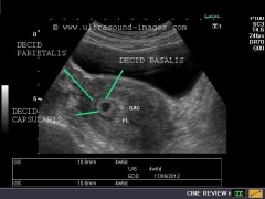
**Double decidual sac sign | **"Double decidual sac" sign | ||
***Two concentric rings (uterine lining and gestation lining) around anechoic gestational sac | ***Two concentric rings (uterine lining and gestation lining) around anechoic gestational sac) | ||
***Highly suggestive of intrauterine pregnancy (before yolk sac or embryo embryo visualized | ***Highly suggestive of intrauterine pregnancy (before yolk sac or embryo embryo visualized | ||
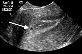
**"Intradecidual sign" | |||
**Intradecidual sign | |||
***Thickened decidua on one side of uterine cavity surrounding anechoeic sac | ***Thickened decidua on one side of uterine cavity surrounding anechoeic sac | ||
| Line 102: | Line 105: | ||
***Definitive evidence of intrauterine pregnancy | ***Definitive evidence of intrauterine pregnancy | ||
***Multiple yolk sacs is earliest sign in multiple gestational pregnancy | ***Multiple yolk sacs is earliest sign in multiple gestational pregnancy | ||
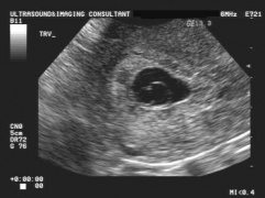
**Double bleb sign | **"Double bleb" sign | ||
***Yolk sac and amniotic cavity that look like two bubbles within gestational sac | ***Yolk sac and amniotic cavity that look like two bubbles within gestational sac | ||
| Line 109: | Line 112: | ||
***First visual manifestation of fetus (or "embryo") | ***First visual manifestation of fetus (or "embryo") | ||
***Thickening of margin on yolk sac | ***Thickening of margin on yolk sac | ||
**Cardiac activity present (FHR ~100-115 bpm) | **Cardiac activity present (FHR ~100-115 bpm) | ||
**Crown rump length: ~5mm | |||
*8-9 weeks | *8-9 weeks | ||
**Limb buds appear | **Limb buds appear | ||
| Line 123: | Line 125: | ||
File:double_decidual_sac_sign.jpg|Double decidual sac sign | File:double_decidual_sac_sign.jpg|Double decidual sac sign | ||
File:intradecidual sign.jpg|Intradecidual sign | File:intradecidual sign.jpg|Intradecidual sign | ||
File:double_bleb_sign.jpg|Double bleb sign (yolk sac and amniotic cavity | File:double_bleb_sign.jpg|Double bleb sign (yolk sac and amniotic cavity) | ||
File:fetal_pole.jpg|Fetal pole (Thickening of yolk sac on one side) | File:fetal_pole.jpg|Fetal pole (Thickening of yolk sac on one side) | ||
</gallery> | </gallery> | ||
| Line 134: | Line 136: | ||
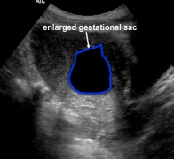
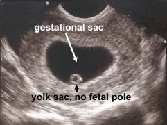
*Gestational sac >18mm without fetal pole | *Gestational sac >18mm without fetal pole | ||
*Collapsed gestational sac | *Collapsed gestational sac | ||
* | *Pseudogestational sac | ||
**Endometrial breakdown during ectopic pregnancy | **Endometrial breakdown during [[ectopic pregnancy]] | ||
**May be erroneously interpreted as true gestational sac in ectopic pregnancy | **May be erroneously interpreted as true gestational sac in ectopic pregnancy | ||
**Irregularly shaped | **Irregularly shaped | ||
| Line 143: | Line 145: | ||
*Absence of fetal heart beat in embryo with CRL >5mm | *Absence of fetal heart beat in embryo with CRL >5mm | ||
*FHR <90 bpm | *FHR <90 bpm | ||
*Molar pregnancy | *[[Molar pregnancy]] | ||
**Many small, irregular cystic structures in endometrium | **Many small, irregular cystic structures in endometrium | ||
**"Snowstorm appearance" | **"Snowstorm appearance" | ||
| Line 152: | Line 154: | ||
File:collapsed gestational sac.jpg|Collapsed gestational sac | File:collapsed gestational sac.jpg|Collapsed gestational sac | ||
File:pseudogestational sac.jpg|Pseudogestational sac | File:pseudogestational sac.jpg|Pseudogestational sac | ||
File:molar pregnancy.jpg|Molar pregnancy with multiple cystic lesions in endometrium | File:molar pregnancy.jpg|Molar pregnancy with multiple cystic lesions in endometrium ("snowstorm appearance") | ||
</gallery> | </gallery> | ||
====Ectopic pregnancy==== | ====[[Ectopic pregnancy]]==== | ||
*Implantation of blastocyst outside of endometrium | *Implantation of blastocyst outside of endometrium | ||
*Occur in fallopian tubes, cervix, ovaries, peritoneal cavity, or scar of prior uterine surgery | *Occur in fallopian tubes, cervix, ovaries, peritoneal cavity, or scar of prior uterine surgery | ||
*Most definitive sonographic sign is gestational sac with yolk sac, embryo, or fetal heart beat outside of the endometrium | *Most definitive sonographic sign is gestational sac with yolk sac, embryo, or fetal heart beat outside of the endometrium | ||
*Tubal | *Tubal pregnancy | ||
** | **"Tubal ring" sign: echogenic thick-walled anechoic sac visualized between uterus and ovary (separate from ovary when pressure applied to fallopian tube with probe) | ||
*Interstitial Pregnancy | *Interstitial Pregnancy | ||
**Implantation of gestational sac in muscular wall of uterus at proximal regional of fallopian tubes | |||
**"Interstitial line" sign: hyperechoic line from endometrial stripe to cornual region adjacent to interstitial gestational sac | |||
*Cervical Pregnancy | *Cervical Pregnancy | ||
**Implantation of gestational sac below internal cervical os | **Implantation of gestational sac below internal cervical os | ||
**Can cause life-threatening hemorrhage | **Can cause life-threatening hemorrhage | ||
**Must distinguish between cervical ectopic and spontaneous abortion at level of cervix: | **Must distinguish between cervical ectopic and spontaneous abortion at level of cervix: | ||
{| {{table}} | |||
| align="center" style="background:#f0f0f0;"|'''Cervical ectopic''' | |||
| align="center" style="background:#f0f0f0;"|'''Spontaneous abortion at cervix''' | |||
|- | |||
| Hour-glass shaped uterus||Elongated gestational sac | |||
|- | |||
| Gestational sac with yolk sac, fetal pole, or heartbeat||Absent embryonic cardiac activity | |||
|- | |||
| Closed internal os||Dilated internal os | |||
|- | |||
| Color doppler showing hypervascular trophoblastic ring||Sac moves in cervix with gentle probing (“Sliding sac sign”) | |||
|- | |||
| Empty uterine cavity||Loss of sac on serial imaging | |||
|- | |||
| | |||
|} | |||
<gallery mode="packed"> | <gallery mode="packed"> | ||
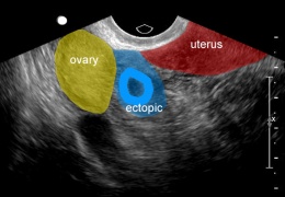
File:ectopic pregnancy.jpg|Ectopic pregnancy | File:ectopic pregnancy.jpg|Ectopic pregnancy | ||
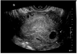
File:free_fluid_ectopic.png|Free fluid in cul-de-sac with no gestational sac visualized in uterus | File:free_fluid_ectopic.png|Free fluid in cul-de-sac with no gestational sac visualized in uterus | ||
File:tubal ring.jpg|Ectopic pregnancy between ovary and uterus, suggestive of tubal ectopic | File:tubal ring.jpg|Ectopic pregnancy between ovary and uterus, suggestive of tubal ectopic | ||
File:cervical ectopic.jpg| | File:interstitial pregnancy.png|Interstitial pregnancy | ||
File:cervical ectopic.jpg|Cervical pregnancy | |||
</gallery> | </gallery> | ||
===Ovarian | ===[[Ovarian Torsion]]=== | ||
* | *Bimodal age distribution: young women (15-30yo) and post-menopausal women | ||
*Typically occur in enlarged ovaries (>5cm) | |||
**Benign mature cystic teratomas | |||
**Hemorrhagic/large ovarian cysts | |||
**Cystic neoplasms | |||
**Cystadenomas | |||
**Polycystic ovarian syndrome | |||
*Sonographic signs associated with torsion | |||
**Enlarged ovary from edema, engorgement, hemorrhage | |||
**Midline ovary | |||
**Free pelvic fluid | |||
**Underlying ovarian lesions | |||
**Peripherally displaced follicular cysts | |||
**Doppler findings: | |||
***Decreased/absent venous or arterial flow | |||
***Absent/reversed diastolic flow | |||
***Can have normal flow (from dual supply from uterine and ovarian arteries) | |||
<gallery mode="packed"> | <gallery mode="packed"> | ||
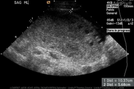
File:simple_cyst.jpg| | File:simple_cyst.jpg|Large ovarian cystadenoma | ||
File:hemorrhagic_cyst.jpg|Hemorrhagic cyst with blood clot | File:hemorrhagic_cyst.jpg|Hemorrhagic ovarian cyst with blood clot | ||
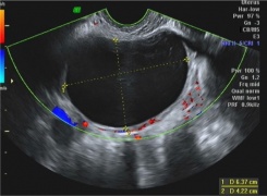
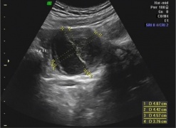
File: ovarian_torsion 1.png|Large midline ovary with multiple peripherally located follicular cysts (torsion) | |||
File:absent_waveforms.png|Absent color doppler and venous and arterial waveforms (torsion) | |||
</gallery> | </gallery> | ||
==Pearls and Pitfalls== | ==Pearls and Pitfalls== | ||
*Free pelvic fluid with positive hCG is ectopic pregnancy until proven otherwise | |||
*Presence of doppler flow cannot rule out ovarian torsion | |||
==Documentation== | ==Documentation== | ||
| Line 204: | Line 241: | ||
==References== | ==References== | ||
<references/> | <references/> | ||
*www.radiopaedia.org | |||
*www.acep.org/sonoguide | |||
*www.fetalultrasound.com | |||
[[Category:Ultrasound]] | [[Category:Ultrasound]] | ||
[[Category:Radiology]] | [[Category:Radiology]] | ||
[[Category:OBGYN]] | |||
Latest revision as of 21:39, 17 December 2019
Background

- Also known as endovaginal ultrasound
- Used to evaluate female reproductive organs including uterus, ovaries, fallopian tubes, cervix, and vagina
Indications
General
- Pelvic pain
- Abnormal pelvic or abdominal exam
Uterus
- Vaginal bleeding
- Fibroids
- IUD placement
Ovaries and adnexa
- Ovarian cyst
- Ovarian torsion
- Ovarian masses
- Tubo-ovarian abscess
- Hydrosalpinx
Pregnancy
- Confirm intrauterine pregnancy
- Gestational dating
- Fetal monitoring and surveillance
- Evaluation of placenta
- Spontaneous abortion
- Intrauterine fetal demise
- Ectopic pregnancy
- Heterotopic pregnancy
- Molar pregnancy
Technique
General
- Use 5 MHz endocavitary probe (high frequency, low penetration)
- Apply surgical lubricant inside and outside probe cover
- Place patient in lithotomy position
- Gently advance probe into vaginal canal and position adjacent to cervix
- May be more comfortable for patient to insert probe into vagina herself
- Apply gentle pressure to lower abdominal wall with one hand and manipulate probe with other hand
Longitudinal scan
- Probe marker pointing at 12:00
- Shows anterior structures on left side of screen
- Begin midline at endometrial stripe
- Scan from right to left through uterus
- Move probe laterally to view adnexa and ovaries
Transverse scan
- Rotate probe 90 degrees counterclockwise so probe marker is pointing at 9:00
- Shows right-sided structures on left side of screen
- Start at endometrial stripe and scan throughout uterus
- Locate cornual flare (junction of uterus and fallopian tubes)
- Move probe laterally along fallopian tube to locate ovary
- Ovaries identified by follicular (anechoic or hypoechoic) structures
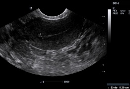
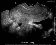
Uterus in longitudinal plane
Cornual flare (uterus and fallopian tube junction) in transverse plane
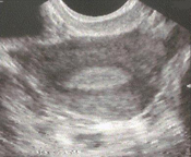
Normal ovary with follicles


Normal Findings
Uterus
- Anteverted: Fundus pointing to anterior abdominal wall
- Retroverted: Fundus pointing to posterior wall
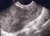
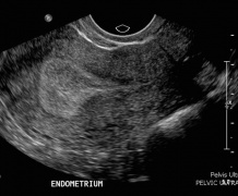
- Endometrial stripe: Measured from thickest echogenic area (from one basal endometrial interface to the other, including canal)
- Pre-menopausal:
- During menstruation: 2-4mm
- Early proliferative phase: 5-7mm
- Late proliferative phase: 11mm
- Secretory phase: 7-16mm
- After D&C or SAB: >5mm
- Consider retained POC if thicker
- Post-menopausal:
- With vaginal bleeding: <5mm
- Without vaginal bleeding: <11mm
- Pre-menopausal:
Normal thickened endometrial stripe during secretory phase
Normal thin endometrial stripe during menses


Ovaries
- Physiologic cysts present
- <3 cm diameter
- Ovarian follicle or corpus luteum
- Typically don't cause complications
Pregnancy (1st trimester)
- 4-5 weeks
- Gestational sac
- First sign of early pregnancy, usually seen between 3-5 weeks gestation
- "Double decidual sac" sign
- Two concentric rings (uterine lining and gestation lining) around anechoic gestational sac)
- Highly suggestive of intrauterine pregnancy (before yolk sac or embryo embryo visualized
- "Intradecidual sign"
- Thickened decidua on one side of uterine cavity surrounding anechoeic sac
- Gestational sac
- 5-6 weeks:
- Yolk Sac
- Circular (or sometimes as two parallel lines) echogenic structure with thick walls within gestational sac
- Definitive evidence of intrauterine pregnancy
- Multiple yolk sacs is earliest sign in multiple gestational pregnancy
- "Double bleb" sign
- Yolk sac and amniotic cavity that look like two bubbles within gestational sac
- Yolk Sac
- 6-7 weeks
- Fetal Pole
- First visual manifestation of fetus (or "embryo")
- Thickening of margin on yolk sac
- Cardiac activity present (FHR ~100-115 bpm)
- Crown rump length: ~5mm
- Fetal Pole
- 8-9 weeks
- Limb buds appear
- Head identifiable
- 9-10 weeks
- FHR ~170-180 bpm
- Fetal movement visible
- End of embryonic period
Double decidual sac sign
Intradecidual sign
Double bleb sign (yolk sac and amniotic cavity)
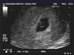
Fetal pole (Thickening of yolk sac on one side)


Abnormal Findings
Abnormal Pregnancy
- Gestational sac >10mm without visible yolk sac
- Gestational sac >18mm without fetal pole
- Collapsed gestational sac
- Pseudogestational sac
- Endometrial breakdown during ectopic pregnancy
- May be erroneously interpreted as true gestational sac in ectopic pregnancy
- Irregularly shaped
- Located in endometrial cavity, instead of eccentrically within endometrium
- No yolk sac present
- May not have double decidual sac sign
- Absence of fetal heart beat in embryo with CRL >5mm
- FHR <90 bpm
- Molar pregnancy
- Many small, irregular cystic structures in endometrium
- "Snowstorm appearance"
Gestational sac greater than 10mm without yolk sac
Gestational sac greater than 18mm without fetal pole
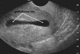
Collapsed gestational sac
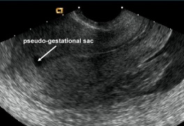
Pseudogestational sac
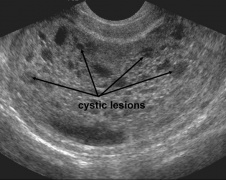
Molar pregnancy with multiple cystic lesions in endometrium ("snowstorm appearance")


Ectopic pregnancy
- Implantation of blastocyst outside of endometrium
- Occur in fallopian tubes, cervix, ovaries, peritoneal cavity, or scar of prior uterine surgery
- Most definitive sonographic sign is gestational sac with yolk sac, embryo, or fetal heart beat outside of the endometrium
- Tubal pregnancy
- "Tubal ring" sign: echogenic thick-walled anechoic sac visualized between uterus and ovary (separate from ovary when pressure applied to fallopian tube with probe)
- Interstitial Pregnancy
- Implantation of gestational sac in muscular wall of uterus at proximal regional of fallopian tubes
- "Interstitial line" sign: hyperechoic line from endometrial stripe to cornual region adjacent to interstitial gestational sac
- Cervical Pregnancy
- Implantation of gestational sac below internal cervical os
- Can cause life-threatening hemorrhage
- Must distinguish between cervical ectopic and spontaneous abortion at level of cervix:
| Cervical ectopic | Spontaneous abortion at cervix |
| Hour-glass shaped uterus | Elongated gestational sac |
| Gestational sac with yolk sac, fetal pole, or heartbeat | Absent embryonic cardiac activity |
| Closed internal os | Dilated internal os |
| Color doppler showing hypervascular trophoblastic ring | Sac moves in cervix with gentle probing (“Sliding sac sign”) |
| Empty uterine cavity | Loss of sac on serial imaging |
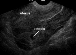
Ectopic pregnancy
Free fluid in cul-de-sac with no gestational sac visualized in uterus
Ectopic pregnancy between ovary and uterus, suggestive of tubal ectopic
Interstitial pregnancy
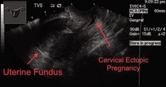
Cervical pregnancy


Ovarian Torsion
- Bimodal age distribution: young women (15-30yo) and post-menopausal women
- Typically occur in enlarged ovaries (>5cm)
- Benign mature cystic teratomas
- Hemorrhagic/large ovarian cysts
- Cystic neoplasms
- Cystadenomas
- Polycystic ovarian syndrome
- Sonographic signs associated with torsion
- Enlarged ovary from edema, engorgement, hemorrhage
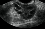
- Midline ovary
- Free pelvic fluid
- Underlying ovarian lesions
- Peripherally displaced follicular cysts
- Doppler findings:
- Decreased/absent venous or arterial flow
- Absent/reversed diastolic flow
- Can have normal flow (from dual supply from uterine and ovarian arteries)
Large ovarian cystadenoma
Hemorrhagic ovarian cyst with blood clot
Large midline ovary with multiple peripherally located follicular cysts (torsion)
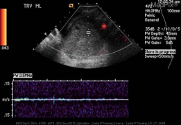
Absent color doppler and venous and arterial waveforms (torsion)


Pearls and Pitfalls
- Free pelvic fluid with positive hCG is ectopic pregnancy until proven otherwise
- Presence of doppler flow cannot rule out ovarian torsion
Documentation
Normal Exam
Abnormal Exam
Clips
External Links
See Also
References
- www.radiopaedia.org
- www.acep.org/sonoguide
- www.fetalultrasound.com