File:MyositisOssificans.png: Difference between revisions
Jstevenson (talk | contribs) (Plain radiograph taken two months after injury demonstrates irregular calcification located along muscle on medial aspect of right thigh. Diagnosis was myositis ossificans.) |
Jstevenson (talk | contribs) No edit summary |
||
| Line 1: | Line 1: | ||
Plain radiograph taken two months after injury demonstrates irregular calcification located along muscle on medial aspect of right thigh. Diagnosis was myositis ossificans. | Plain radiograph taken two months after injury demonstrates irregular calcification located along muscle on medial aspect of right thigh. Diagnosis was myositis ossificans. Kim SY, Park JS, Ryu KN, Jin W, Park SY. Various tumor-mimicking lesions in the musculoskeletal system: causes and diagnostic approach. Korean J Radiol (2011) | ||
Latest revision as of 01:14, 30 August 2016
Plain radiograph taken two months after injury demonstrates irregular calcification located along muscle on medial aspect of right thigh. Diagnosis was myositis ossificans. Kim SY, Park JS, Ryu KN, Jin W, Park SY. Various tumor-mimicking lesions in the musculoskeletal system: causes and diagnostic approach. Korean J Radiol (2011)
File history
Click on a date/time to view the file as it appeared at that time.
| Date/Time | Thumbnail | Dimensions | User | Comment | |
|---|---|---|---|---|---|
| current | 01:11, 30 August 2016 | 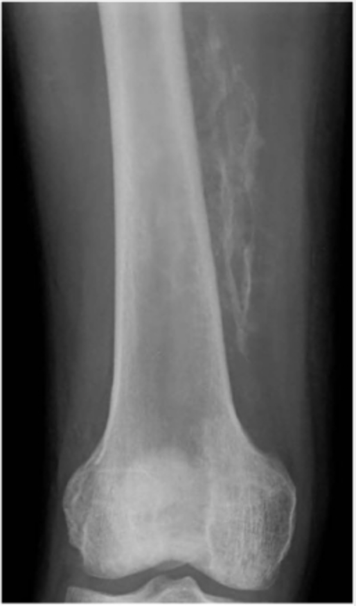 | 514 × 874 (286 KB) | Jstevenson (talk | contribs) | Plain radiograph taken two months after injury demonstrates irregular calcification located along muscle on medial aspect of right thigh. Diagnosis was myositis ossificans. |
You cannot overwrite this file.
File usage
The following page uses this file: