CT brain interpretation: Difference between revisions
No edit summary |
|||
| (10 intermediate revisions by 2 users not shown) | |||
| Line 1: | Line 1: | ||
==Mnemonic== | ==Mnemonic== | ||
[[File:Brainlobes.png|thumb|Lobes of the brain.]] | |||
[[File:Cranial bones en.png|thumb|Bones of the cranium.]] | |||
[[File:Computed tomography of human brain - large.png|thumb|Example of a normal head CT with IV contrast.]] | |||
''Blood Can Be Very Bad'' | ''Blood Can Be Very Bad'' | ||
*Blood | *Blood | ||
| Line 17: | Line 20: | ||
**Blood becomes hypodense at 2wks (exact time depends on size of clot) | **Blood becomes hypodense at 2wks (exact time depends on size of clot) | ||
*Findings | *Findings | ||
**Epidural | **[[Epidural hematoma]] (blood problem) | ||
***Lens shaped | ***Lens shaped | ||
***Does not cross sutures | ***Does not cross sutures | ||
***Classically described with injury to middle meningeal artery | ***Classically described with injury to middle meningeal artery | ||
***Low mortality if treated prior to unconsciousness (<20% morbidity/mortality) | ***Low mortality if treated prior to unconsciousness (<20% morbidity/mortality) | ||
**Subdural (brain problem) | **[[SDH|Subdural]] (brain problem) | ||
***Sickle shaped | ***Sickle shaped | ||
***Crosses sutures but not midline | ***Crosses sutures but not midline | ||
| Line 31: | Line 34: | ||
****Suprasellar cistern is first place see SAH (location adjacent to circle of willis) | ****Suprasellar cistern is first place see SAH (location adjacent to circle of willis) | ||
***Aneurysm: 80% | ***Aneurysm: 80% | ||
***AVM: 5% | ***[[AVM]]: 5% | ||
**Intraventricular/Intraparenchymal Hemorrhage | **Intraventricular/Intraparenchymal Hemorrhage | ||
***Typically obvious findings | ***Typically obvious findings | ||
| Line 56: | Line 59: | ||
*Look at gyral pattern all the way around (gyri effacement indicator of increased ICP) | *Look at gyral pattern all the way around (gyri effacement indicator of increased ICP) | ||
*Findings | *Findings | ||
**Tumor | **[[brain tumor|Tumor]] | ||
***May see increased hypodensity (edema) | ***May see increased hypodensity ([[cerebral edema in brain cancer|edema]]) | ||
***80% | ***80% visible without contrast | ||
**Atrophy | **Atrophy | ||
**Abscess | **[[brain abscess|Abscess]] | ||
**Hemorrhagic contusion | **Hemorrhagic contusion | ||
**Mass effect | **Mass effect | ||
**Stroke | **[[Stroke]] | ||
**Intracranial air (skull fracture) | **Intracranial air ([[skull fracture]]) | ||
**Hyperdense middle cerebral artery or basilar artery sign | **Hyperdense middle cerebral artery or basilar artery sign | ||
***Suggests thrombosis of vessel | ***Suggests thrombosis of vessel | ||
**Suggestive of [[Cerebral venous thrombosis]] : | **Suggestive of [[Cerebral venous thrombosis]] : | ||
***Empty delta sign: dense triangle in superior sagittal sinus | ***Empty delta sign: dense triangle in superior sagittal sinus | ||
***Cord sign: hyperattenuated, | ***Cord sign: hyperattenuated, homogeneous linear or round foci in cerebral sinus <ref>Ram K. P. Vijay. "The Cord Sign."Radiology. 2006; 240:299-300.</ref> | ||
***Vein Sign: hypoattenuated foci in the deep vein <ref>Linn J, Pfefferkorn T. "Noncontrast CT in Deep Cerebral Venous Thrombosis and Sinus Thrombosis: Comparison of Its Diagnostic Value for Both Entities."AJNR Am J Neuroradiology. 2010; 30: 728-735. </ref> | ***Vein Sign: hypoattenuated foci in the deep vein <ref>Linn J, Pfefferkorn T. "Noncontrast CT in Deep Cerebral Venous Thrombosis and Sinus Thrombosis: Comparison of Its Diagnostic Value for Both Entities."AJNR Am J Neuroradiology. 2010; 30: 728-735. </ref> | ||
| Line 75: | Line 78: | ||
*Check all 4 for size and for hemorrhage | *Check all 4 for size and for hemorrhage | ||
**Temporal tips (comma-shaped) of lateral ventricle first place to show hydrocephalus | **Temporal tips (comma-shaped) of lateral ventricle first place to show hydrocephalus | ||
**If enlarged must differentiate between hydrocephalus from increased pressure versus atrophy: | **If enlarged must differentiate between [[hydrocephalus]] from increased pressure versus atrophy: | ||
***Are the gyri effaced? If yes suggestive of increased pressure | ***Are the gyri effaced? If yes suggestive of increased pressure | ||
==Bone== | ==Bone== | ||
*Inspect petrous ridges for skull base fracture | *Inspect petrous ridges for [[basilar skull fracture|skull base fracture]] | ||
*Look at mastoid air cells full of fluid (blood) for indirect evidence of fracture | *Look at mastoid air cells full of fluid (blood) for indirect evidence of fracture | ||
==Examples== | |||
<gallery mode="packed"> | |||
File:Intracerebral heamorrage.jpg|[[Hemorrhagic stroke]] (spontaneous intracranial hemorrhage) | |||
File:Epidural Hematoma.jpg|[[Epidural hemorrhage]] | |||
File:Subduralandherniation.png|[[Subdural hemorrhage]] | |||
File:SubarachnoidP.png|[[Subarachnoid hemorrhage]] | |||
File:Hydrocephalus (cropped).jpg|[[Hydrocephalus]] | |||
File:PMC4857327 10.1177 2050313X15591314-fig3.png|[[Brain abscess]] | |||
File:StrokeMCA overlay.png|[[Ischemic stroke]] | |||
</gallery> | |||
==See Also== | ==See Also== | ||
Latest revision as of 22:29, 25 January 2023
Mnemonic


Blood Can Be Very Bad
- Blood
- Cisterns
- Brain
- Ventricles
- Bone
Blood
- Questions
- Is blood present?
- If so, where is it?
- If so, what effect is it having?
- Physiology
- Acute blood is bright white (once it clots)
- Blood becomes isodense at 1wk (exact time depends on size of clot)
- Blood becomes hypodense at 2wks (exact time depends on size of clot)
- Findings
- Epidural hematoma (blood problem)
- Lens shaped
- Does not cross sutures
- Classically described with injury to middle meningeal artery
- Low mortality if treated prior to unconsciousness (<20% morbidity/mortality)
- Subdural (brain problem)
- Sickle shaped
- Crosses sutures but not midline
- Marker for severe head injury (mortality approaches 80%)
- Small amount of bleed can be associated with major shift (secondary to brain injury/oozing)
- SAH
- Blood in the cisterns/cortical gyral surface/interhemispheric fissure
- Suprasellar cistern is first place see SAH (location adjacent to circle of willis)
- Aneurysm: 80%
- AVM: 5%
- Blood in the cisterns/cortical gyral surface/interhemispheric fissure
- Intraventricular/Intraparenchymal Hemorrhage
- Typically obvious findings
- Unimportant if intraventricular ruptured into parenchyma or vice-versa
- Epidural hematoma (blood problem)
Cisterns
- 4 key cisterns:
- Circummesencephalic
- First cistern to show increased ICP (squished shut)
- Suprasellar
- Quadrigeminal
- "W" shaped
- Second cistern to show increased ICP
- Sylvian
- May see isloated distal MCA bleed
- Circummesencephalic
- 2 questions:
- Is there blood?
- Are the cisterns open?
Brain
- Compare side to side
- Look for grey-white differentiation
- Grey is denser so appears lighter on CT
- Look at gyral pattern all the way around (gyri effacement indicator of increased ICP)
- Findings
- Tumor
- May see increased hypodensity (edema)
- 80% visible without contrast
- Atrophy
- Abscess
- Hemorrhagic contusion
- Mass effect
- Stroke
- Intracranial air (skull fracture)
- Hyperdense middle cerebral artery or basilar artery sign
- Suggests thrombosis of vessel
- Suggestive of Cerebral venous thrombosis :
- Tumor
Ventricles
- Check all 4 for size and for hemorrhage
- Temporal tips (comma-shaped) of lateral ventricle first place to show hydrocephalus
- If enlarged must differentiate between hydrocephalus from increased pressure versus atrophy:
- Are the gyri effaced? If yes suggestive of increased pressure
Bone
- Inspect petrous ridges for skull base fracture
- Look at mastoid air cells full of fluid (blood) for indirect evidence of fracture
Examples
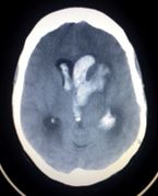
Hemorrhagic stroke (spontaneous intracranial hemorrhage)


.jpg)


See Also
- Head CT (Clinical Decision Rules)
- CT Before Lumbar Puncture
- Intracerebral Hemorrhage (ICH)
- X-ray interpretation (main)
References
- Blood Can Be Very Bad: CT Interpretation Course Guide
- www.uic.edu/com/ferne/pdf/acep2005_spring/perron_acep2005_spring_bcbvb_course.pdf