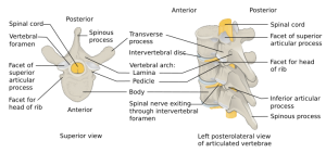
Jefferson fracture
Background
- Also known as a C1 burst fracture
- No ligamentous disruption
- Is an unstable spine injury
Vertebral fractures and dislocations types
- Cervical fractures and dislocations
- Thoracic and lumbar fractures and dislocations
Clinical Features
- Fracture of the anteterior and posterior arches[1]
- Due to axial loading transmitted through occipital condyles to the lateral masses
- Frequently associated with other cervical fractures
- May be associated with vertebral artery injury[2]
Differential Diagnosis
Neck Trauma
- Penetrating neck trauma
- Blunt neck trauma
- Cervical injury
- Neurogenic shock
- Spinal cord injury
Evaluation
- Suspect disruption if:
- Lateral x-ray: Increase in the predental space between C1 and dens (>3mm in adults, >5mm in children)
- Odontoid x-ray: Masses of C1 lie lateral to outer margins of articular pillars of C2
- If either of the above findings on x-ray obtain CT C-spine
Management
Prehospital Immobilization
Hospital
- Degree of instability determined by whether or not the transverse ligament is disrupted
- C-collar
- Consult ortho or spine as needed
Disposition
- Admit
See Also
References
- ↑ Jefferson, G. (1919) ‘Fracture of the atlas vertebra. Report of four cases, and a review of those previously recorded’, British Journal of Surgery, 7(27), pp. 407–422.
- ↑ Muratsu H, Doita M, Yanagi T et-al. Cerebellar infarction resulting from vertebral artery occlusion associated with a Jefferson fracture. J Spinal Disord Tech. 2005;18 (3): 293-6.


