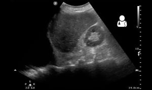
Hemothorax
Background
- Each hemithorax can hold 40% of circulating blood volume
Clinical Features
- Diminished or absent breath sounds
- Dullness to percussion
Differential Diagnosis
Thoracic Trauma
- Airway/Pulmonary
- Cardiac/Vascular
- Musculoskeletal
- Other
Evaluation

CXR with large right sided hemothorax (and widened mediastinum).
- CXR
- Upright - Fluid collections >200-300cc can usually be seen
- Supine - Fluid collections >1000cc can be missed
- Mainstem bronchus intubation can appear like a hemothorax on CXR
- US
- CT is gold standard
Management
- Tube thoracostomy
- Evacuation of 1000-1500mL of blood immediately or 200mL/hr x 4hr = consider thoracotomy
- Autotransfuse lost blood if possible
Adult Chest Tube Sizes
| Chest Tube Size | Type of Patient | Underlying Causes |
| Small (8-14 Fr) |
|
|
| Medium (20-28 Fr) |
|
|
| Large (36-40 Fr) |
|
Disposition
- Admit


