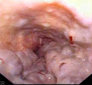
File:Esophageal varices - wale.jpg
No higher resolution available.
Esophageal_varices_-_wale.jpg (316 × 293 pixels, file size: 17 KB, MIME type: image/jpeg)


File history
Click on a date/time to view the file as it appeared at that time.
You cannot overwrite this file.
File usage
The following 2 pages use this file: