Rectal foreign body
Background
- Most are in the rectal ampulla and therefore palpable on digital examination
- Make sure that object is not sharp before exam
- Injuries may consist of hematoma, lacerations (w/ potential perforation)
Clinical Presentation
- Rectal pain and/or fullness
- History of rectal foreign body placement
Differential Diagnosis
Anorectal Disorders
- Anal fissure
- Anal fistula
- Anal malignancy
- Anal tags
- Anorectal abscess
- Colorectal malignancy
- Condyloma acuminata
- Constipation
- Crohn's disease
- Cryptitis
- GC/Chlamydia
- Hemorrhoids
- Pedunculated polyp
- Pilonidal cyst
- Proctitis
- Pruritus ani
- Rectal foreign body
- Rectal prolapse
- Syphilitic fissure
Diagnosis
- Abdominal xray
- Demonstrate position, shapes, and number of foreign bodies
- Demonstrate position, shapes, and number of foreign bodies
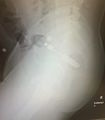
Foreign body noted in rectum on lateral abdominal xr

- Demonstrates possible presence of free air (perforation of rectum or colon)
- Perf of rectum below peritoneal reflection shows extraperitoneal air along psoas
- Perf above peritoneal reflection reveals intraperitoneal free air under diaphragm
- Demonstrates possible presence of free air (perforation of rectum or colon)
- CT
- Useful when foreign body is radiolucent and for detection of free air
Management
ED removal
Suitable for non-sharp objects that are in the distal rectum
- IV sedation and analgesia usually needed for adequate relaxation for removal of larger FB's
- Local anesthesia (perianal block) will relax the anal sphincter and may be needed.
- Anal lubrication
- In lithotomy position, suprapubic pressure with DRE and valsalva may deliver object without instrumentation.
- If obstetric forceps needed, pt should bear down as object is extracted.
- Large bulbar objects create a vacuum-like effect
- Break vacuum by passing foley behind object, inject air and pull foley out (balloon up)
- Observe for at least 12hr to ensure that object did not perforate the rectum
Surgical Consultation Indications
- Removal could injure the sphincter
- ED attempts fail
- Risk of ischemia, perforation, or if excess manipulation required (risk of bacteremia)
See Also
Source
- Tintinalli
- Roberts