Clavicle fracture: Difference between revisions
| Line 22: | Line 22: | ||
==Evaluation== | ==Evaluation== | ||
[[File:Clavicle Fracture Left.jpg|thumb|Left clavicle fracture on xray.]] | |||
*Assess distal pulse, motor, and sensation | *Assess distal pulse, motor, and sensation | ||
*X-ray | *X-ray | ||
Revision as of 15:27, 13 June 2020
This page is for adult patients; see Clavicle fracture (peds) for pediatric patients
Background
- Generally secondary to shoulder trauma (direct trauma over the clavicle is less common)
- Fractured segment:
- Middle third: 80%
- Distal third: 15%
- Medial third: 5%
- Distal fracture may be associated with coracoclavicular ligament rupture
- Medial fracture may be associated with intrathoracic injuries
Clinical Features
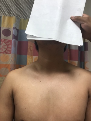
- Swelling, deformity, and tenderness overlying the clavicle
- Affected arm may be supported by the contralateral arm
Differential Diagnosis
Thoracic Trauma
- Airway/Pulmonary
- Cardiac/Vascular
- Musculoskeletal
- Other
Shoulder and Upper Arm Diagnoses
Traumatic/Acute:
- Shoulder Dislocation
- Clavicle fracture
- Humerus fracture
- Scapula fracture
- Acromioclavicular joint injury
- Glenohumeral instability
- Rotator cuff tear
- Biceps tendon rupture
- Triceps tendon rupture
- Septic joint
Nontraumatic/Chronic:
- Rotator cuff tear
- Impingement syndrome
- Calcific tendinitis
- Adhesive capsulitis
- Biceps tendinitis
- Subacromial bursitis
- Cervical radiculopathy
Refered pain & non-orthopedic causes:
- Referred pain from
- Neck
- Diaphragm (e.g. gallbladder disease)
- Brachial plexus injury
- Axillary artery thrombosis
- Thoracic outlet syndrome
- Subclavian steal syndrome
- Pancoast tumor
- Myocardial infarction
- Pneumonia
- Pulmonary embolism
Evaluation
- Assess distal pulse, motor, and sensation
- X-ray
- May be seen on chest x-ray, shoulder x-ray, or dedicated clavicle films (preferred)
- If high suspicion and no fracture on plain films, consider CT
Management
- Place the affected extremity in a sling
- Pain management
Disposition
- Almost all may be discharged with orthopedic surgery follow-up
- Urgent follow-up indicated for (possible need for surgical intervention):
- Displacement
- Comminution
- >2cm of shortening
- Orthopedic surgery consultation in the ED for:
- Skin tenting
- Open fracture
- Neurovascular compromise


