File:Periodontium.png
Original file (339 × 665 pixels, file size: 128 KB, MIME type: image/png)
Summary
Download original file 339 × 665 px svg View in browser You need to attribute the author
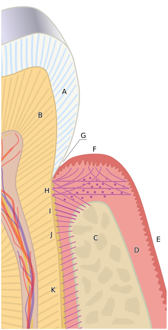
By Goran tek-en, CC BY-SA 4.0, https://commons.wikimedia.org/w/index.php?curid=30709759 A diagram of the periodontium. The crown of the tooth is covered by enamel (A). Dentin (B). The root of the tooth is covered by cementum. C, alveolar bone. D, subepithelial connective tissue. E, oral epithelium. F, free gingival margin. G, gingival sulcus. H, principal gingival fibers. I, alveolar crest fibers of the periodontal ligament (PDL). J, horizontal fibers of the PDL. K, oblique fibers of the PDL. Goran tek-en The Periodontium
CC BY-SA 4.0 File:Periodontium.svg Created: 20 January 2014 About this interface | Discussion | Help
File history
Click on a date/time to view the file as it appeared at that time.
| Date/Time | Thumbnail | Dimensions | User | Comment | |
|---|---|---|---|---|---|
| current | 20:43, 2 December 2021 | | 339 × 665 (128 KB) | Rossdonaldson1 (talk | contribs) | Download original file 339 × 665 px svg View in browser You need to attribute the author By Goran tek-en, CC BY-SA 4.0, https://commons.wikimedia.org/w/index.php?curid=30709759 A diagram of the periodontium. The crown of the tooth is covered by ename... |
You cannot overwrite this file.
File usage
The following 4 pages use this file: