File:Hip fracture classification.png
Original file (1,335 × 1,272 pixels, file size: 301 KB, MIME type: image/png)
Summary
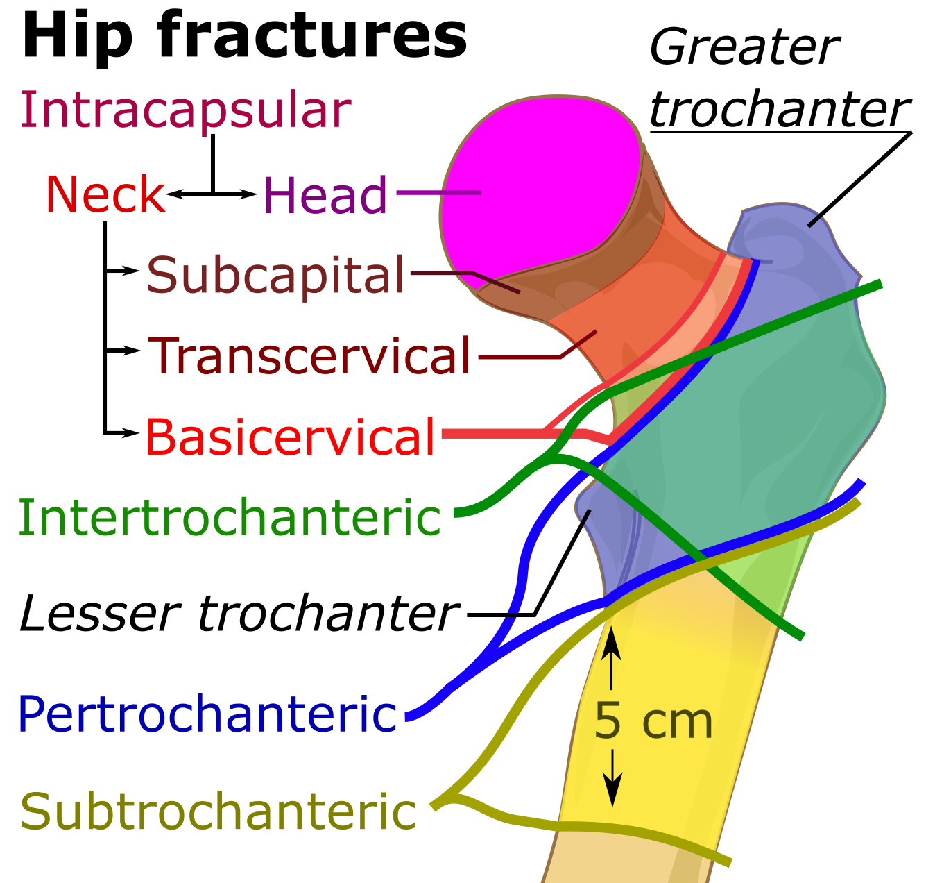
By Mikael Häggström, using image by Mariana Ruiz Villarreal (LadyofHats - Source image: File:Human skeleton front en.svg by Mariana Ruiz Villarreal (LadyofHats). Public DomainSources for areas:Prevention and Management of Hip Fracture on Older People. Section 7: Surgical management. Scottish Intercollegiate Guidelines Network. Retrieved on 2017-04-23. Last modified 15/7/02Area of trochanteric fractures: Ernst Raaymakers, Inger Schipper, Rogier Simmermacher, Chris van der Werken. Proximal femur. AO Foundation. Retrieved on 2017-04-23.Area of subtrochanteric fractures: Mark A Lee. Subtrochanteric Hip Fractures. Retrieved on 2017-04-25. Updated: Jun 22, 2016Area of femoral neck fractures: Page 333 in: Paul Tornetta, III, Sam W. Wiesel (2010) Operative Techniques in Orthopaedic Trauma Surgery, Lippincott Williams & Wilkins ISBN: 9781451102604.edit, CC0, https://commons.wikimedia.org/w/index.php?curid=58330229
File history
Click on a date/time to view the file as it appeared at that time.
| Date/Time | Thumbnail | Dimensions | User | Comment | |
|---|---|---|---|---|---|
| current | 22:37, 17 March 2021 | | 1,335 × 1,272 (301 KB) | Rossdonaldson1 (talk | contribs) | By Mikael Häggström, using image by Mariana Ruiz Villarreal (LadyofHats - Source image: File:Human skeleton front en.svg by Mariana Ruiz Villarreal (LadyofHats). Public DomainSources for areas:Prevention and Management of Hip Fracture on Older People... |
You cannot overwrite this file.
File usage
The following 6 pages use this file: