Talus fracture
Revision as of 04:34, 4 January 2014 by Rossdonaldson1 (talk | contribs) (Created page with "==Hindfoot== ===Talus=== ====Background==== *Almost always associated with other injuries ====Diagnosis==== *CT often required for accurate diagnosis ====Management==== *Maj...")
Hindfoot
Talus
Background
- Almost always associated with other injuries
Diagnosis
- CT often required for accurate diagnosis
Management
- Major fracture (talar neck and head)
- Immediate ortho consultation required (high rate of avascular necrosis)
- Minor fracture
- Posterior splint, NWB, ortho referral
Calcaneus
Background
- Associated injuries are common
- Types
- Intra-articular (75%)
- Sclerotic line may be only evidence of impacted fracture
- Extra-articular (25%)
- Anterior process fx is most common
- Intra-articular (75%)
Diagnosis
- Imaging
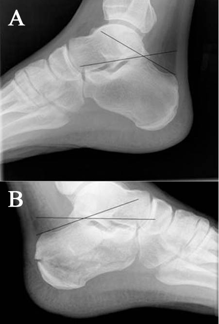
- Decreased Boehler's angle (<25') may be only sign of fx (compare w/ opposite side)
Treatment
- Intra-articular fracture
- Immobilization w/ posterior splint
- Non-weightbearing
- Elevation (very important - fx has high rate of severe swelling)
- Ortho consult
- Extra-articular fracture
- Immobilization and close ortho f/u
Images
- (A) Normal Boehler's angle and (B) Abnormal Boehler's angle
Midfoot
LisFranc Injury
- See Lisfranc Injury
- All are diagnosed/managed in similar way
- Imaging: (weight-bearing AP, lateral, oblique)
- CT sometimes necessary
- Treatment: Non-weightbearing short leg cast, ortho referral
- Imaging: (weight-bearing AP, lateral, oblique)
Forefoot
Fifth Metatarsal
Background
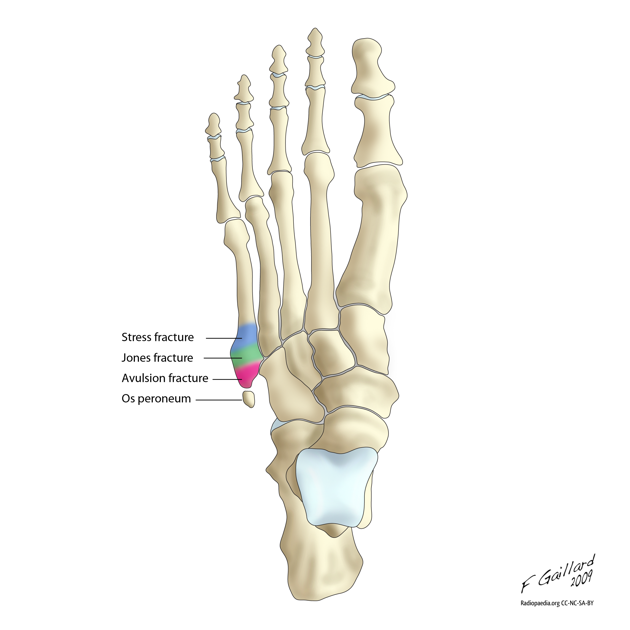
- Os peroneum is an accessory bone (ossicle) located at the lateral side of the tarsal cuboid, proximal to the base of 5th metatarsal, commonly mistaken for fx
3 types of 5th metatarsal fx:
- Tuberosity (styloid) avulsion fracture:
- Most common fx at base of 5th metatarsal
- Sx often mild, pts usually present with sprained ankle complaint
- Occurs due to forced inversion foot/ankle while in plantar flexion
- Jones or metaphyseal-diaphyseal junction fracture:
- Second most common fx at base of 5th metatarsal
- Abrupt onset of lateral foot pain, with no prior h/o pain at that site, suggests acute injury and helps distinguish from stress injury
- Occurs due to sudden change in direction w/ heel off the ground
- Edema & ecchymosis usually present, may not be able to bear weight
- Diaphyseal stress fracture:
- Occurs through repetitive microtrauma, usually in younger athletes
- Important to identify given propensity for delayed union and nonunion
- Usually present with h/o months of pain, which is more intense during exercise or weight-bearing
- always ask about persistent pain prior to acute event to help distinguish worsening stress fx from acute fx
Diagnosis
Plain radiographs are usually adequate
- Must distinguish Jones fx from diaphyseal stress freacture:
- Acute fx will have narrow fx line that appears sharp, normal thin cortex adjacent to fx, and normal intramedullary canal
- Stress fx will demonstrate cortical thickening near fx line, older stress fx will demonstrate widened fx line and intramedullary sclerosis
Management
- Tuberosity (Styloid) Avulsion Fracture
- Refer to ortho if > 3mm displacement
- Nondisplaced fx usually require only symptomatic tx, RICE
- Walking boot (casting rarely necessary) and weight-bearing as tolerated, f/u in 1 week
- Jones Fracture (non-displaced)
- Posterior splinting, strict NWB, RICE, ortho f/u in 3-5 days
- 50% of Jones fx treated conservatively may result in nonunion or refracture
- Conservative tx failure usually due to poor vascular supply of bone and premature return to weight-bearing
- Diaphyseal Stress Fracture
- Strict NWB short-leg cast, RICE
- Ortho referral for all stress fxs
Metatarsal
Background
- Must rule-out associated Lisfranc injury
Management
- Posterior splint, NWB, ortho referral in 2-3d
Phalange
- Management: buddy-taping, hard-soled shoe
See Also
Source
- Tintinalli
- Uptodate
- Ilustration by Dr. Frank Gaillard; CC SA NC BY licence
- http://radiopaedia.org/articles/jones_fracture


