Knee x-rays: Difference between revisions
No edit summary |
|||
| (2 intermediate revisions by one other user not shown) | |||
| Line 6: | Line 6: | ||
==Examples== | ==Examples== | ||
<gallery mode="packed"> | <gallery mode="packed"> | ||
File:Lipohemarthrosis.png|Lipohemarthrosis with subtle [[tibial plateau fracture]] | |||
File:Patellaluxation ap 001.png|[[Patella dislocation]] | File:Patellaluxation ap 001.png|[[Patella dislocation]] | ||
File:Fracpetella.png|[[Patella fracture]] lateral | File:Fracpetella.png|[[Patella fracture]] lateral | ||
File:Patella fracture.jpg|[[Patella fracture]] AP | File:Patella fracture.jpg|[[Patella fracture]] AP | ||
File:Segond.jpg|[[Segond fracture]] | |||
File:TibPlateauBadMark.png|[[Tibial plateau fracture]] | |||
File:TibPlatFracPlainMark.png|[[Tibial plateau fracture]] | |||
</gallery> | </gallery> | ||
| Line 19: | Line 23: | ||
==References== | ==References== | ||
<references/> | <references/> | ||
[[Category:Orthopedics]] | |||
Latest revision as of 14:18, 16 November 2017
Background
Technique
Knee x-rays
- Anteroposterior and lateral views
- Consider sunrise if pain over patella
Examples
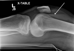
Lipohemarthrosis with subtle tibial plateau fracture

Patella fracture lateral


