Ocular ultrasound: Difference between revisions
(link placements) |
Neil.m.young (talk | contribs) (SN and SP added) |
||
| Line 22: | Line 22: | ||
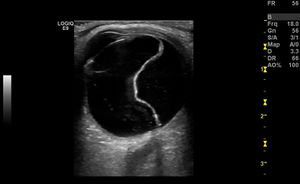
*Echogenic undulating membrane in the posterior globe, protruding into the vitreous | *Echogenic undulating membrane in the posterior globe, protruding into the vitreous | ||
*Evaluate with patient moving eye left/right | *Evaluate with patient moving eye left/right | ||
*SN 97-100% and SP 83-100%<ref>Vrablik, ME, et al. The Diagnostic Accuracy of Bedside Ocular Ultrasonography for the Diagnosis of Retinal Detachment: A Systematic Review and Meta-analysis. Annals of Emergency Medicine. 2015; 65(2):199–203.e1.</ref> | |||
[[File:Retinal detachment.jpg|thumb|Retinal detachment ultrasound]] | [[File:Retinal detachment.jpg|thumb|Retinal detachment ultrasound]] | ||
Revision as of 02:24, 5 February 2015
Technique
- Use vascular/linear probe
- Plenty of ultrasound gel to decrease amount of pressure needed to place on eye
Elevated ICP
- Measure optic nerve 3mm posterior to the globe, from inner wall to inner wall
- Normal is <5mm
Globe Rupture
- Only perform if you can ensure that you do not put pressure on the globe
- Findings
- Decrease in size of globe
- Anterior chamber collapse
- Vitreous hemorrhage
- Buckling of the sclera
- see Globe Rupture
Intraocular Foreign Body
- Bright, echogenic acoustic profile w/ associated shadowing or reverberation
Retinal Detachment
- Echogenic undulating membrane in the posterior globe, protruding into the vitreous
- Evaluate with patient moving eye left/right
- SN 97-100% and SP 83-100%[1]
Vitreous Hemorrhage
- Vitreous filled with multiple large echoes
- Increasing the gain is helpful for detecting acute hemorrhages
See Also
Source
Sonoguide
- ↑ Vrablik, ME, et al. The Diagnostic Accuracy of Bedside Ocular Ultrasonography for the Diagnosis of Retinal Detachment: A Systematic Review and Meta-analysis. Annals of Emergency Medicine. 2015; 65(2):199–203.e1.


